
Microscopy Meets Materials

For centuries scientists have used microscopes to explore the hidden world of small dimensions. Today the science of microscopy is an essential link between understanding of structures and properties of materials and tailoring them for real world applications.

Accordingly, our main research activities are devoted to developing new microscopic characterisation methods, functional nanostructures, and the practical applications of advanced microscopy.

The ability of modern transmission electron microscopes to investigate solids at atomic resolution has led to a tremendous progress in materials science. With the basis of a monochromated transmission electron microscope (TEM) and an atomic resolution STEM (ASTEM), the groups of Werner Grogger, Gerald Kothleitner and Ferdinand Hofer concentrate on the development of nanoanalytical methods using EELS and EDXS. These advanced methods are used to explore the structure of materials and devices as diverse as transistors, turbine blades and biomaterials.h.

Our major research activities are:

Methodical developments
Nanotomography
Atomic resolution STEM of nanostructures
Nanoparticles and carbon materials
EELS-imaging of surface plasmons

The controlled fabrication of functional structures with feature sizes down to the nanoscale can be considered as a central gateway for current but in particular for upcoming applications in science and technology. Within that broad research field, additive direct-write manufacturing became increasingly relevant during the past decade[[1]], as that technology pool allows for unique applications, including the particular aspect of true 3D printing[[2]]. To pave the way for new developments, a basic understanding of physical and chemical processes together with technological aspects is indispensably needed.

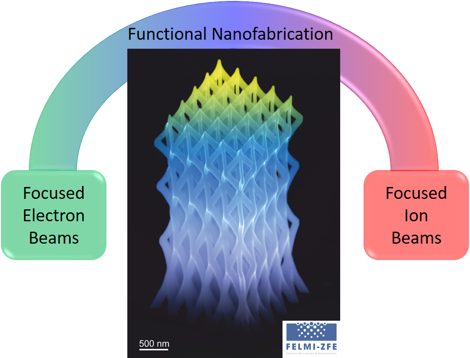
Based on that awareness, this work-area focuses on the fundamental understanding of novel approaches with the aspiration to derive new concepts for real applications in science and technology[[3]]. While strongest focus is currently placed on 3D Nano-Printing (3DNP) via Focused Electron Beam Induced Deposition (FEBID)[[4]], we also use Focused Ion Beam (FIB) processing for additive and subtractive (nano)fabrication.

Aside of the scientific orientation, we work together with industry in the frame of the Christian Doppler Laboratory for Direct-Write Fabrication of 3D Nano-Probes (CDL-DEFINE), where 3DNP is used for research and development of novel application concepts for atomic force microscopy nano-probes.

Research and development in highest-performance materials requires increasingly sophisticated investigation methods. Dynamic experiments both in scanning and transmission electron microscopy enable the investigation of the progress of physical processes and chemical reactions on the micro- and nanoscale. Correlations between the microstructure of a material, its composition and its behaviour under various stresses (mechanical, thermal …) can be deduced. Another advantage of in situ experiments is the fact that materials can be investigated under conditions they experience in their practical applications. Subsequent 3D reconstructions of the bulk of the material will provide additional information about the material’s properties and behaviour. Correlation of the results is gained from both the dynamic experiments and the 3D reconstructions which will facilitate the design and development of new materials.

3D reconstruction of the separation layer of a microfiltration membrane.

IFELMI-ZFE has a long tradition of cutting edge research in the field of real-time and in situ characterisation of bio-degradable and bio-compatible materials. During the past decades these materials have gained more and more importance due to environmental protection and medical sciences issues. Current core activities are 3D investigations of neuronal networks, microfiltration membranes and crack propagation in polymers. In situ characterisation of enzymatic cellulose degradation in real-time as well as in situ testing of membranes and polymers have become major research areas. 3D sectioning / imaging / reconstruction aspects under static, dynamic and even cryogenic conditions allow us to get a comprehensive insight into material properties at the micro- and nanoscale. Against this challenging background we are driven by the desire to move beyond current limitations.

3D reconstruction (scale bar: 5 µm) of part of a neuron of the neural network active in the processing of signals from a locust eye using serial blockface scanning electron microscopy (SBEM) and showing the distribution of the afferent synaptic connections (yellow).
[Cooperation with the Medical University of Graz]

Single enzyme imaging (arrows) on cellulose surfaces via AFM high-resolution imaging in liquid conditions (a) cross confirmed via material sensitive phase imaging (b). This method also allows dynamic real-time investigations in liquid environments revealing enzymatic activities with site-specific (amorphous / crystalline) information (c).