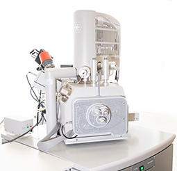
The FEI ESEM Quanta 600 FEG is a versatile scanning electron microscope with three imaging modes. The “high vaccum mode” (HV) is a conventional SEM mode with the need of conventional specimen preparation. In the “low vacuum mode” (LV) electrically non conductive samples can be imaged without the need of a conductive layer (e.g. carbon, gold etc.). Additionally in the “ESEM mode” (ESEM) wet samples can be investigated in their “natural” state. The thermally assisted field emission gun (FEG) delivers high brightness of the electron beam and high imaging resolution. Additionally the microcroscope can be equipped with a tensile stage, a Peltier cooled specimen stage, a heating stage and an in situ ultramicrotome.
FEI ESEM Quanta 600 FEG
Environmental Scanning Electron Microscope
Key Features
- Seamless „point and click“ transition between imaging modes
- Superior low vacuum, low kV imaging simultaneous secondary electron (SE) and backscattered electron (BSE) imaging in LV mode
- Allows for in situ dynamic experiments
- True surface (SE) imaging in all vacuum modes and voltages
- Easy-to-use, four quadrant/single quadrant user interface
Essential Specifications
Resolution
• < 2.0 nm @ 30 kV SE @ HV
• < 2.0 nm @ 30 kV SE @ ESEM
• < 3.5 nm @ 3 kV SE @ LV
• < 1.5 nm @ 30 kV STEM @ HV
Emitter
Thermal Field Emission Gun (FEG)
Accelerating Voltage
0.2 – 30 kV
Probe Current
Can be measured externly with a Faraday cup
Detectors
• Everhart Thornley Detector (ETD): SE, BSE @ HV
• Large Field Detector (LFD): SE @ LV
• Solid State Backscattered Electron Detector SSD-BSD:BSE @ HV, LV
• Gaseous Secondary Electron Detector(GSED):SE @ESEM
• EDS Detector Thermo Noran Vantage
5-Axes Motorised Eucentric Specimen Stage
• X = 150 mm / Y = 150 mm / Z = 60 mm
• Rotation = 360° (continuous)
• Tilt = -5° – +70°
Image Processing
• Resolution: up to 3584 x 3094 pixel
• Dwell: 100 ns – 1 ms per pixel
System Control
• Windows 2000™ based 32-bit graphical user interface
Additional methods
Deben tensile stage MT5000
Peltier cooled specimen stage
Heating-stage
In situ ultramicrotomy