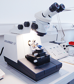
Ultramicrotomy (including cryoultramicrotomy) is a standard method for the preparation of ultrathin/semi-thin sections as well as very flat surfaces of plastics, biological and biomedical objects for various microscopic investigations.
Improvements in preparation techniques over the last few decades have demonstrated that thin sections of different materials that are free from artefacts can be successfully prepared for electron microscopic investigations.
Advantages Ultramicrotomy:
- favorable & fast technique
- Clean technique, no contamination, no smearing
- No beam damage
- No amorphisation or implanation
Typical Applications of Microtomy:
- Polymers (Laminates, blends, additives, etc.)
- Material Investigation (Inclusions, contaminations, surface defects, identification)
- Fiber and foil investigations
- Analysis of hard & soft materials
- Biological investigations
Cryo-Ultramicrotomy
enables to make thin sections of constant thickness with very soft materials that cannot be cut at ambient temperature. The material is brought close to transition temperature Tg (if known) to obtain a harder consistency. For all cryo sectioning of biological as well as industrial specimens, the use of an ionizer is essential.
Cryo-ultramicrotomy requires a lot of practise and is highly time-consuming.
Examples:
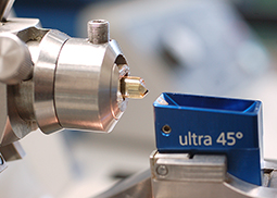
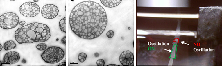
Ultra-sonic knife:
New demands for the block-face smoothness have led to the development of an ultra-sonic oscillating knife (Diatome, Switzerland). It offers the advantage to section materials at room temperature, which normally require cryo temperatures. This development leads to thinner sections without compression and significantly better structure preservation.
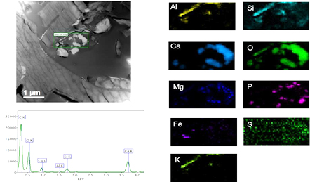
Analytical scanning transmission electron microscopy:
Pieces of biochar were covered in gold all-around in the Leica EM ACE 600 (Leica Microsystems, Vienna, Austria; 45 mA, 8,0 × 10⁻³ mbar, working distance 50 mm) using a rotating device to enable 3D coating. This resulted in a minimum of 25-nm-thick gold layer on the biochar. These nuggets were embedded in Spezifix40 (Struers, Willich, Germany) and after hardening carefully trimmed with a trim 90 blade (Diatome, Switzerland).
The preparation of 50 nm slices was done at room temperature with the ultra-sonic-knife (Ultra Sonic, Diatome) for reducing compression and allowing best structure preservation in the ultramicrotome Leica EM UC6 (Leica Microsystems).